[ad_1]
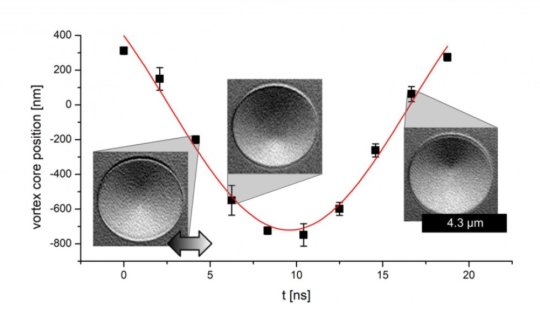
In the future, today’s electronic storage technology may be superseded by devices based on tiny magnetic structures. These individual magnetic regions correspond to bits and need to be as small as possible and capable of rapid switching. In order to better understand the underlying physics and to optimize the components, various techniques can be used to visualize the magnetization behavior. Scientists at Johannes Gutenberg University Mainz (JGU) in Germany have now refined an electron microscope-based technique that makes it possible not only to capture static images of these components but also to film the high-speed switching processes. They have also employed a specialized signal processing technology that suppresses image noise.
“This provides us with an excellent opportunity to investigate magnetization in small devices,” Daniel Schönke of the JGU Institute of Physics explained. The research was carried out in cooperation with Surface Concept GmbH and the results have been published in the journal Review of Scientific Instruments.
Scanning electron microscopy with polarization analysis is a lab-based technique for imaging magnetic structures. Compared with optical methods, it has the advantage of high spatial resolution. The main disadvantage is the time it takes to acquire an image in order to achieve a good signal-to-noise ratio. However, the time required to measure the periodically excited and therefore periodically changing magnetic signal can be shortened by using a digital phase-sensitive rectifier that only detects signals of the same frequency as the excitation.
Such signal processing requires measurements to be time-resolved. The instrumentation developed by the scientists at JGU provides a time resolution of better than 2 nanoseconds. As a result, the technique can be employed to investigate high-speed magnetic switching processes. It also makes it possible to both capture images and select individual images at a defined point in time within the entire excitation phase.
New technique compares favorably with more complex imaging techniques
This development means the technique is now comparable with the much more complex imaging techniques used at large accelerator facilities and opens up the possibility of investigating the magnetization dynamics of small magnetic components in the laboratory.
The research was carried out within the framework of the Collaborative Research Center CRC/Transregio 173 “Spin+X: Spin in its collective environment,” which is based at Johannes Gutenberg University Mainz and TU Kaiserslautern and financed by the German Research Foundation (DFG). The CRC/TRR involves interdisciplinary teams of researchers from the fields of chemistry, physics, mechanical engineering, and process engineering, who undertake research into magnetic effects with a view to converting these into applications. The primary focus is on the phenomenon of spin. Physicists use this term to refer to the intrinsic angular momentum of a quantum particle, such as an electron or proton. This underlies many magnetic effects.
The development of the novel technique results from the successful and close collaboration of the researchers with the company Surface Concept GmbH, a spin-off of Johannes Gutenberg University Mainz.
Story Source:
Materials provided by Johannes Gutenberg Universitaet Mainz. Note: Content may be edited for style and length.
[ad_2]


